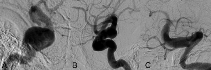
Fig 4.
An 87-year-old man with a history of a right third-nerve palsy. A, Right ICA cerebral angiogram demonstrates a 20-mm cavernous carotid fusiform aneurysm with associated dilation of the supraclinoid ICA as well. B, Left ICA cerebral angiogram shows dilation of the left supraclinoid ICA to approximately 10 mm. C, Left vertebral artery cerebral angiogram shows diffuse dilation and tortuosity of the basilar artery measuring 9 mm in maximum diameter. The cause of the third-nerve palsy was thought to be the right cavernous aneurysm.

