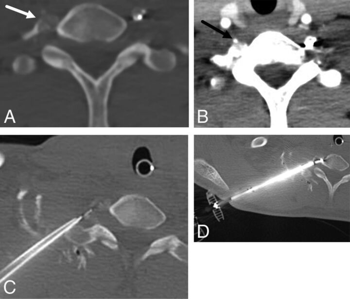
Fig 2.
A 14-year-old boy with painful right transverse process C7 osteoid osteoma (A, arrow). Preprocedural neck CT angiography demonstrates the course and location of the right vertebral artery (B, arrow). C, Thermal monitoring and protection are achieved by placement of a thermocouple and spinal needle in the right C7–T1 neuroforamen. D, RF ablation is performed by using a bipolar navigational probe with slight posterior articulation of the probe tip for optimal positioning.

