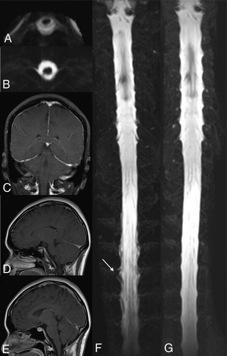
Fig 3.

A 34-year-old man with orthostatic headache. CSF leakage at the spine with epidural fluid accumulation (A) and CSF signals along the neural sleeve (B) were seen at the patient's initial MRM. DPE (C), venous engorgement (D), and pituitary hyperemia (E) were noted at the initial brain MR imaging. 3D maximum intensity projection of the initial MRM (F) revealed an irregular contour along the neural sleeves at the T-spine, indicating CSF leakage (arrow) and reduced CSF volumes with lower CSF intensities of the dural sac compared with the 3D MIP of his recovery MRM (G).
