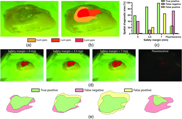
Fig. 5.
(a) Photograph of the tumor mimicking fluorescent optical tissue phantom with regions of varying PPIX concentration. (b) Photograph of the tumor mimicking fluorescent optical tissue phantom with overlaid map of PPIX concentration regions. (c) Margin delineation accuracy for the tumor mimicking fluorescent optical tissue phantom via fluorescence-guided Raman spectroscopic margin delineation with safety margins of 0, 3.5, and 7 mm, and via fluorescence imaging. (d) Margin delineation of tumor mimicking optical tissue phantom using fluorescence-guided Raman spectroscopic margin delineation (with safety margins of 0, 3.5, and 7 mm) and fluorescence imaging. (e) Corresponding true positive, false negative, and false positive areas for fluorescence-guided Raman spectroscopic margin delineation (with safety margins of 0, 3.5, and 7 mm) and fluorescence imaging. Note that safety margins for the probe-tracking system are dependent on both the probe tracking accuracy and the diagnostic model accuracy.

