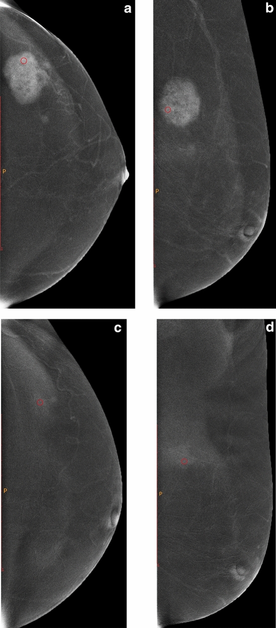
Figure 1.

Female, 51 years old, with lesion located in the outer upper quadrant of the left breast. (a,b) are the subtraction images of CC and MLO views of CESM before NAC, and (c,d) are the subtraction images of CC and MLO views of CESM after two cycles of NAC, respectively. The CC and MLO view grey value reduction percentages were 40.27% and 16.13%, respectively. Before surgery, core needle biopsy confirmed that the patient had invasive breast cancer. After surgical resection, the pathological results showed no invasive cancer components with MP Grade 5.
