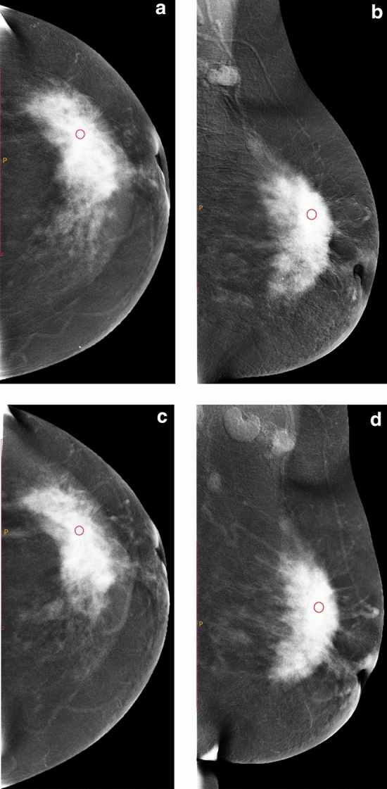
Figure 2.

Female, 49 years old, with lesion located in the left outer quadrant. a and b are the subtraction images of CC and MLO views of CESM before NAC, and c and d are the subtraction images of CC and MLO views of CESM after two cycles of NAC, respectively. The CC and MLO view grey value reduction percentages were 4.02% and 4.95%, respectively. Before surgery, core needle biopsy confirmed that the patient had invasive breast cancer. After surgical resection, the pathological results showed invasive ductal carcinoma Grade II with MP Grade 3.
