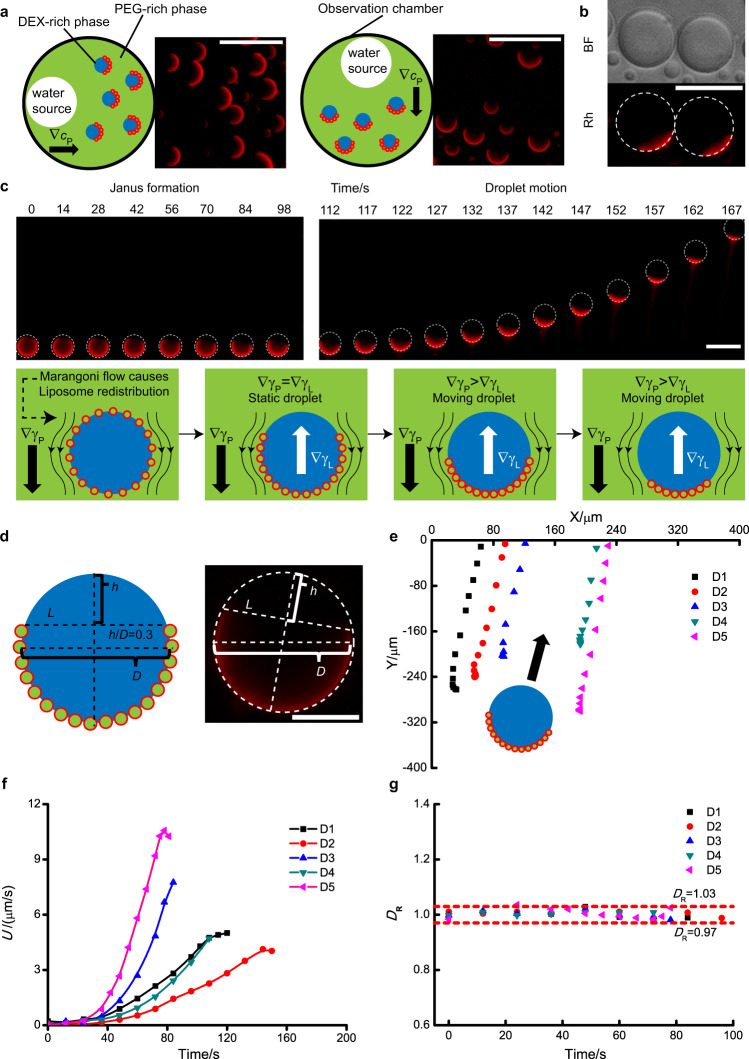
Fig. 3. Formation of Janus droplets and subsequent droplet motion.
a Janus conformation aligns with the position of water addition. Pictures were extracted from Supplementary Movies 1 and 2 and cropped for better presentation. is the polymer gradient. b Droplet structure under bright field (BF) or Rhodamine fluorescence channel (Rh). c Janus formation and droplet motion. In respond to water gradient, the droplet gradually transformed into a Janus droplet. The morphology change under the rhodamine channel is due to the redistribution/desorption of liposomes since Fig. 3b shows that droplets stay spherical under bright field despite the Janus morphology under the rhodamine channel. When the ratio of h to D reached ∼0.3, the droplet starts a directional motion towards the water source, with a characteristic tail of liposomes due to liposome desorption from the droplet surface during the motion. ∇γP and ∇γL are the IFT gradients caused by polymer gradient and liposomes, respectively. Frames were extracted from Supplementary Movie 1 and cropped for better presentation. d Illustration for the Janus droplet with h/D = 0.3. The concentric circle and the line L were drawn as auxiliary lines, and then a line h that is perpendicular to the line L was drawn. The diameter of the concentric circle and the length of line is D and h, respectively. e Trajectories of moving droplets. D1–D5 are representative droplets chosen from Supplementary Movie 1. Arrow corresponds to the direction of movement. f Corresponding velocity (U) and g diameter change (DR, the ratio of droplet diameter at different time points to its average diameter during motion) for droplets in (d). The time point when directional motion starts were chosen as time zero for each droplet. Scale bar is 50 μm for (a–c) and 20 μm for (d).

