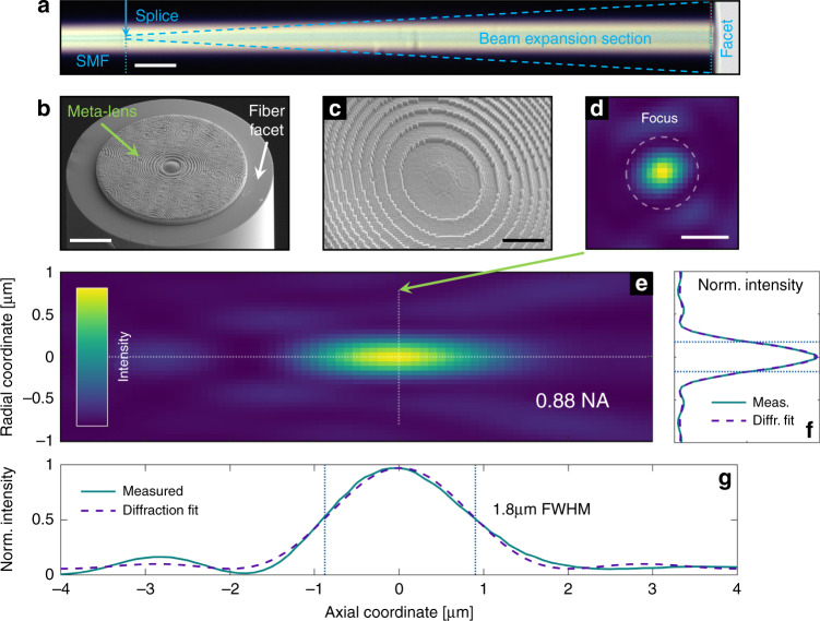
Fig. 3. Measured focusing performance of the implemented UNM-enhanced meta-fibre (sample 2, λ0 = 660 nm in water).
a Microscope image of a functionalized meta-fibre consisting of a single-mode fibre and spliced beam expansion section (scale bar 50 µm). b, c Scanning electron micrographs of an example implemented device (scale bars at 25 µm and 10 µm, respectively). d Measured cross-section of the beam taken at the focal plane (z = 0, scale bar: 500 nm). The dashed circle describes the width of a fitted Airy function. e Axial stack for measured beam cross-sections in cylindrical coordinates obtained by transforming each 2D image into radial symmetry via azimuthal averaging (note that the scale of both axes is the same as that in (d) and (g)). f Radial and (g) axial intensity profiles (solid) along the symmetry axes of the focus, with the dashed lines showing the corresponding fits. In all contour plots, the colour scale ranges linearly from zero (dark blue) to unity (yellow)

