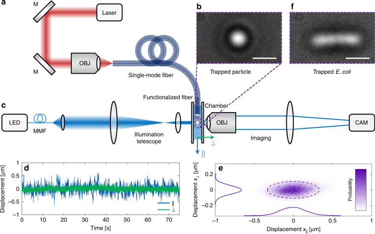
Fig. 4. Optical trapping using a single UNM-enhanced SMF.
a Sketch of the trapping setup consisting of a high-power laser (λ0 = 660 nm) coupled to an UNM-fibre sample, (b) windowed chamber with micro-objects immersed in water, and (c) Koehler illumination (left, a wavelength of 455 nm) and imaging system (right) used to record the motion of the trapped object. The top right images show micrograph examples of (b) a trapped 2 µm silica sphere and (f) a trapped Escherichia coli (E. coli) bacterium (scale bars: 2 µm). d Examples of spatial dynamic displacements for a trapped silica bead along with the transverse (green) and longitudinal (blue) directions for a trapping time of >1 min. e Corresponding 2D representation of the probability with projections on the respective axes. The dashed ellipse depicts the 2σ-environment containing 95% of the points

