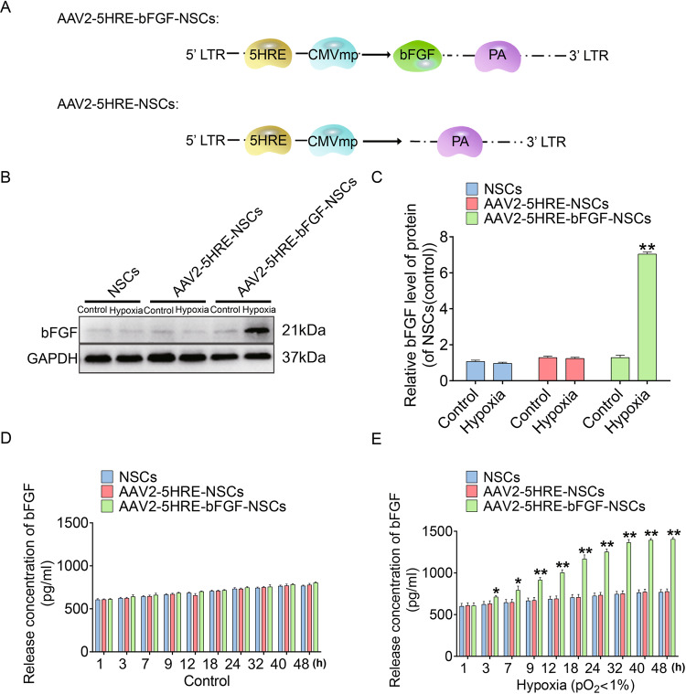
Fig. 1. Preparation and characterization of the AAV2-5HRE-bFGF-NSCs.
A Schematic diagram illustrating the construction of expression vectors AAV2-5HRE and AAV2-5HRE-bFGF. B Western blotting analyses of primary NSCs, AAV2-5HRE-NSCs, and AAV2-5HRE-bFGF-NSCs showing the expression of bFGF under normoxic or hypoxic conditions for 24 h. C The quantitative analyses of bFGF protein expression by western blot. D ELISA assay results showing bFGF secretion in primary NSCs, AAV2-5HRE-NSCs, and AAV2-5HRE-bFGF-NSCs under normoxic conditions. E ELISA assay results showing bFGF secretion of primary NSCs, AAV2-5HRE-NSCs, and AAV2-5HRE-bFGF-NSCs under hypoxic conditions. “*” and “**” represents P < 0.05 or P < 0.01 versus normoxia control. Data are the mean values ± SEM.

