
Figure 4.
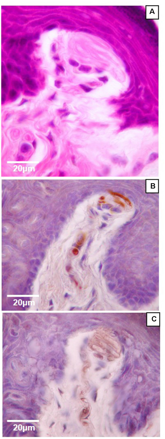
High magnification (100×) light microscopy images of Meissner-like corpuscles in the glabrous footpad. Sagittal sections (8 μm) stained with hematoxylin and eosin (A) or immunostained for neurofilament-200 (B) or S100 (C) and counterstained with hematoxylin (B,C).