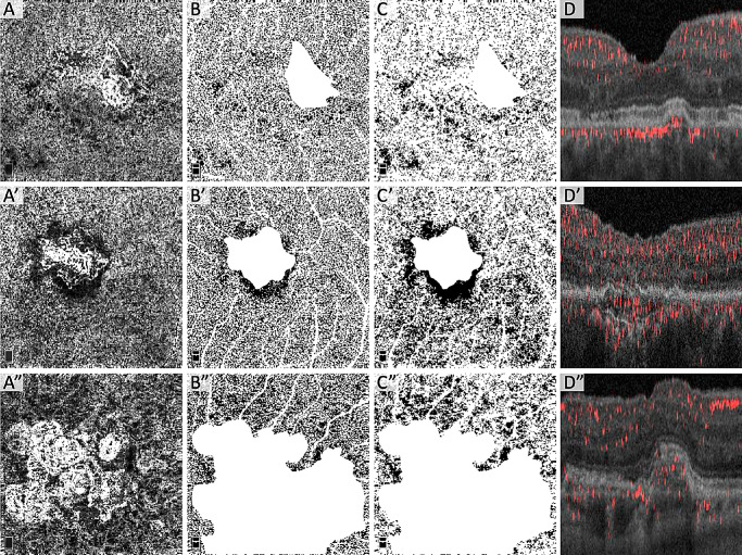
Figure 4.
Choriocapillaris FD stratified by number of CNV flow layers. Each row shows images from the same eye with either one (A–D), two (A’–D’), or three (A”–D”) CNV flow layers. (Column A) Compensated and normalized en face OCTA of the choriocapillaris. (Column B) Local Phansalkar binarized choriocapillaris OCTA of area outside the CNV. (B’) Underestimation of the FD in dark halo region using Phansalkar. (Column C) Global MinError(I) binarized choriocapillaris OCTA of area outside the CNV. (Column D) Cross-sectional projection-resolved OCTA of CNV showing number of CNV flow layers and CNV architecture.

