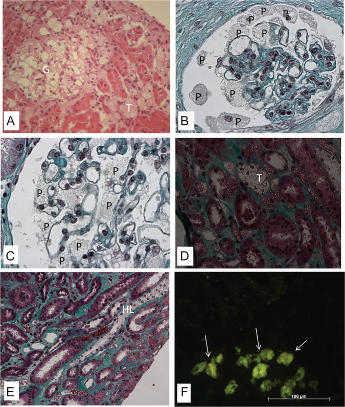
Figure 1.
Kidney biopsy specimen under light microscopy (A-E—embedded in paraffin): The vacuoles seen in the cytoplasm of different cells: especially podocytes (P) in the glomerulus (G), distal tubular cells (T), and Henle loops (HL). (A) Hematoxylin and eosin (magnification: 20× objective; 2.0× optovar). (B-E) Masson’s trichrome (magnification: 160×). (F) Yellowish green natural fluorescence of GL3 in the glomerular and tubular cells (arrows) in frozen section under fluorescence microscopy (magnification: 40× objective; 1.25× optovar).

