Figure 3.
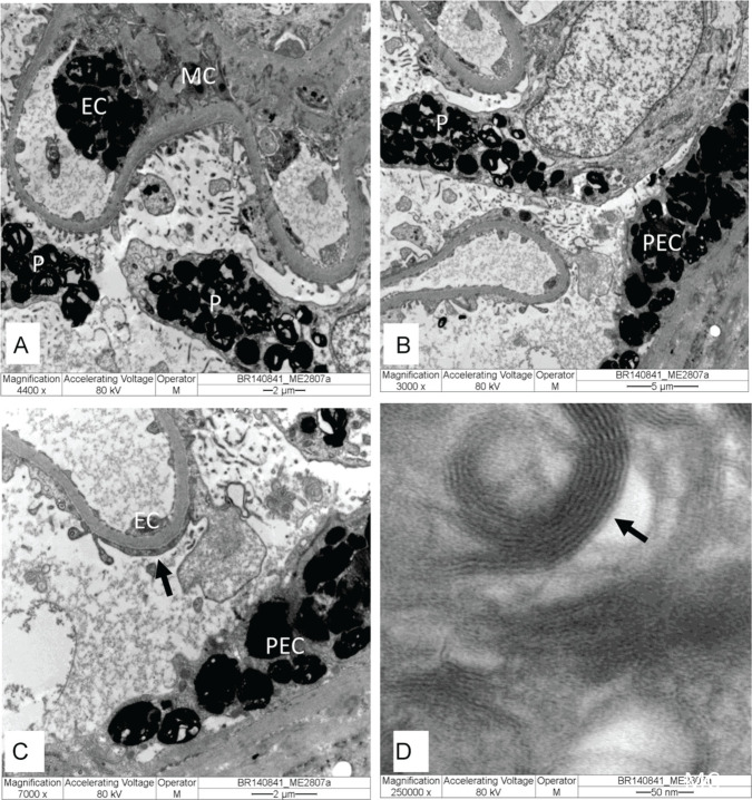
Kidney biopsy specimen under electron microscopy: (A-C) Glomerulus with electron-dense GL3 deposits in endothelial cells (EC), mesangial cells (MC), parietal epithelial cells (PEC), and podocytes (P), with effacement foot process (arrow). (D) In high magnification, GL3 deposits consist of electron-dense multilamellated concentric layers, with periodicity 3.5 to 5 nm (arrow). Original magnification: (A) = 4.400×, (B) = 3.000×, (C) = 7.000×, and (D) = 250.000×.

