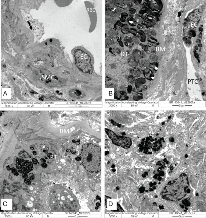
Figure 4.
Kidney biopsy specimen under electron microscopy with electron-dense GL3 deposits in lysosomes in different cells: (A) arteriole in endothelial cells (EC) and smooth muscle cells (SMC); (B) distal tubule (DT) and in endothelial cells (EC) of peritubular capillary (PTC); (C) proximal tubule (PT); and (D) cells in the interstitial compartment. Original magnification: (A-D) = 3.000×.
Note. RBC = red blood cell; BM = basal membrane.

