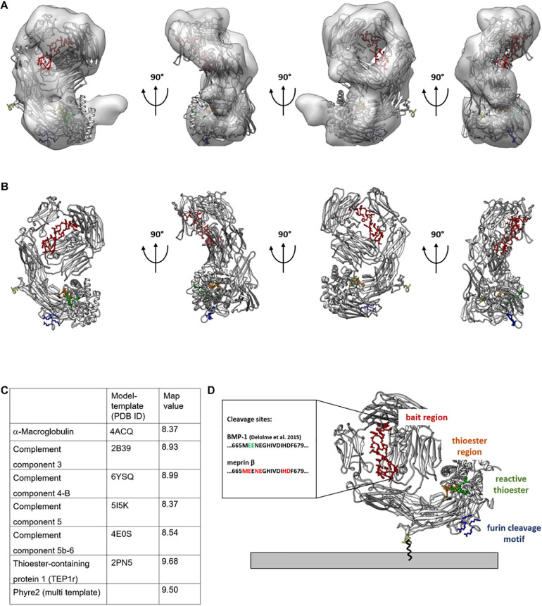
FIGURE 3.
Molecular model of TEP1 fits best into 3D reconstruction of CD109. Thioester-containing protein 1 (TEP1r, PDB ID: 2PN5) matches best with the negative stain transmission electron microscopy (TEM)-generated 3D model. 2PN5 homology model is outlined within the 3D model (A) or by itself (B) rotated in 90° angles with colored functional regions; legend shown in panel (D). For quality reasons, map values were calculated and displayed in tabular view in panel (C). C-terminus expressed in yellow gives an idea of the structural orientation of CD109 on the cell surface (D) Connected to the GPI anchor. The bait region is shown in red with possible cleavage sites given for meprin β and BMP-1; the thioester region is in orange, and its structural proximity to the reactive thioester-defining hexapeptide motif is in green; the furin cleavage site oriented toward the cell membrane is in blue.

