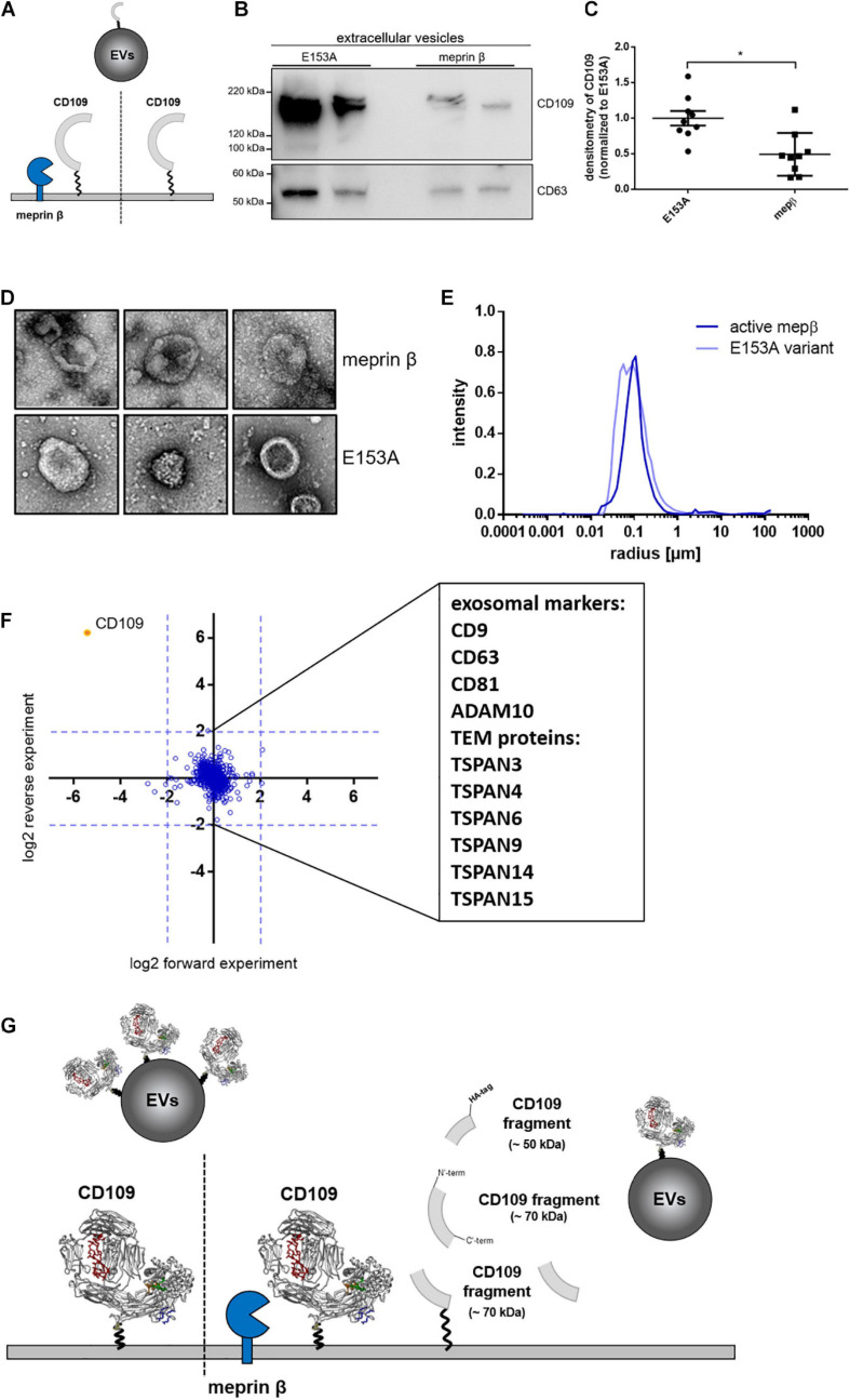
FIGURE 4.
Amount of CD109 on extracellular vesicles reduced after meprin β cleavage. To determine differences of extracellular vesicle (EV) composition between active meprin β and its inactive variant E153A after transient transfection of N-term HA-tagged CD109 stable HEK293T cells [schematic view in panel (A)], an exosome purification kit was used to extract EVs from cell supernatant. Representative Western blot (n = 9) is shown in panel (B) and quantification to exosome marker CD63 (as internal standard control) and afterward normalized to inactive meprin β control (C); statistical analysis was performed using Mann–Whitney test, p < 0.05 was considered statistically significant, *p < 0.01. In panels (D–F), extracellular vesicles were extracted via ultracentrifugation and analyzed in negative stain transmission electron microscopy for size and shape (D) as well as dynamic light scattering analyses; results show no differences in mean radius distribution (E). In the proteomics approach, many exosome markers and tetraspanin-enriched microdomain (TEM) proteins could be detected, but no distinct protein could be detected up- or downregulated besides CD109 (F). In summary, (G) gives a schematic overview of meprin β influencing CD109 not only on the cell surface but also on extracellular vesicle transport.

