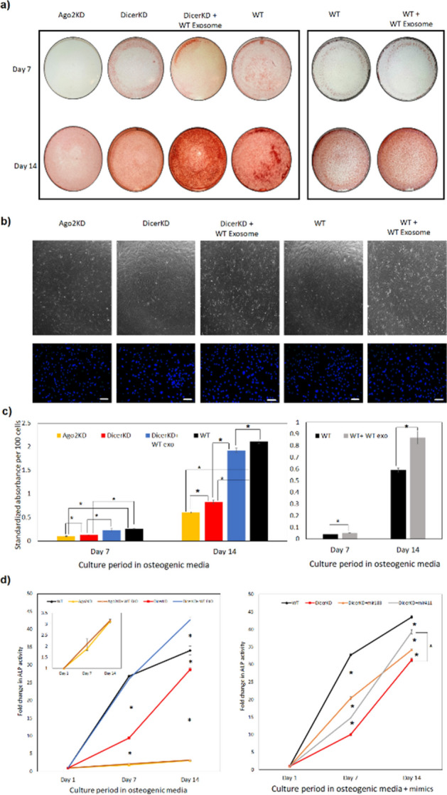
Figure 3.

Differentiation experiments. (a) Alizarin Red staining demonstrate impaired calcium deposition in AGO2KD cells after 7 and 14 days of culture in osteogenic medium. DicerKD cells produced less calcium deposits compared to WT hMSCs at day 7 and 14. Complementation of DicerKD and WT hMSCs with WT exosomes considerably improved calcium deposition. (b) Brightfield microscopic images of experimental groups at day 7 captured at 4 × magnification demonstrated similar confluency level. DAPI staining showed similar number of nuclei per field among different groups. White scale bars are equal to 100 µm. (c) Quantitation of Alizarin Red staining revealed a statistically significant (*P < 0.01) difference among experimental groups. (d) ALP activity was measured using 45 µg of protein from each sample and calculated as standardized fold change at day 7 and 14 compared to day 1 in each group. ALP activity was significantly lower (*P < 0.01) in AGO2KD and DicerKD groups compared to WT hMSCs at day 7 and 14 in osteogenic medium. Complementation of DicerKD hMSCs with WT exosomes restored ALP activity at day 7, and increased ALP activity significantly (*P < 0.01) at day 14 compared to WT hMSCs. Complementation of AGO2KD hMSCs with WT exosomes did not significantly change ALP activity at days 7 and 14 (insert: magnified plot of ALP activity at day 7 and 14 for AGO2KD hMSCs). The addition of miR-183-5p and miR-411-5p mimics to DicerKD cells significantly (*P < 0.01) increased ALP activity at day 7 and 14. Fold changes in ALP activity was calculated compared to day 1 within each group. One-way ANOVA and Tukey's post-hoc test was used for all statistical analyses.
