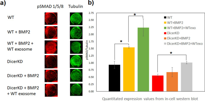
Figure 5.
SMAD1/5/8 Phosphorylation in response to rhBMP2 treatment. (a) Representative images of individual wells showing phosphorylated SMAD1/5/8 staining (red) and corresponding tubulin expression (green) (n = 3 per each group). (b) Quantitation of the images revealed that SMAD 1/5/8 phosphorylation was significantly impaired (*P < 0.01) in DicerKD hMSCs in the presence of BMP2 compared to WT hMSCs. Complementation of DicerKD cells with WT exosomes significantly increased (*P < 0.01) the phosphorylation of SMAD 1/5/8 four hours post rhBMP2 treatment. While these levels were statistically insignificant with respect to WT cells, (P > 0.05), the mean value remained lower indicating a partial restoration of BMP2 function with WT exosome complementation. One-way ANOVA and Tukey's post-hoc test was used for statistical analyses.

