
Figure 8. Rhes levels increase as neurons age can be prevented by Tau mutation.
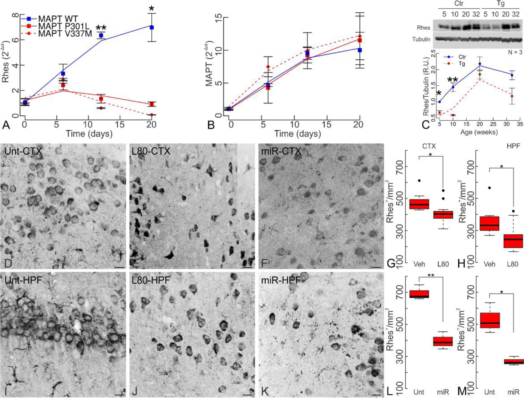
(A) Rhes expression is reduced in MAPT-P301L and MAPT-V337M derived neurons during hiPSC neuronal differentiation as early as the neurorosette stage but increases during cell differentiation in MAPT-WT cells. (B) MAPT levels increase continuously during neuronal differentiation regardless of MAPT genotype. (C) Rhes quantification in rTg4510 transgenic mice (Tg) and littermate controls (Ctr). While Rhes levels increase as mice age in both transgenic and control mice, these are significantly lower in younger transgenic mice (two-way ANOVA, age p=3.24x10⁻³; genotype p=8.01x10⁻³; interaction p=0.88 n.s.). (D-M) Rhes levels reduction in 20 week-old transgenic mice by both lonafarnib and Rhes-miR treatments. Untreated rTg4510 mice had a mean of 693.28±26.92 Rhes+ cells/mm2 in the cortex (CTX) and 529.96±54.91 cells/mm2 in the hippocampus (HPF). Chronic administration of lonafarnib reduced Rhes to 409.09±18.46 cells/mm2 (p=0.018) in the CTX and to 252.55±23.65 cells/mm2 (p=0.019) in the HPF. Rhes-miR reduced Rhes to 396.27±31.52 cells/mm2 (p=0.002) in CTX and to 268.10±16.07 cells/mm2 (p=0.033) in HPF. Statistics shown for Welch’s T tests at *p<0.05, and **p<0.01. (A-C) N=3 (female:male ratio; Ctrs 5 wk 1:2, 10 wk 0:3, 20 wk 2:1, 32 wk 2:1; Tg 5 wk 3:0, 10 wk 0:3, 20 wk 2:1, 32 wk 1:2). (D-M) (female:male ratio; Unt 0:3; L80 3:3; Veh 3:3; Rhes-miR 1:2). Scale bar 100 μm.