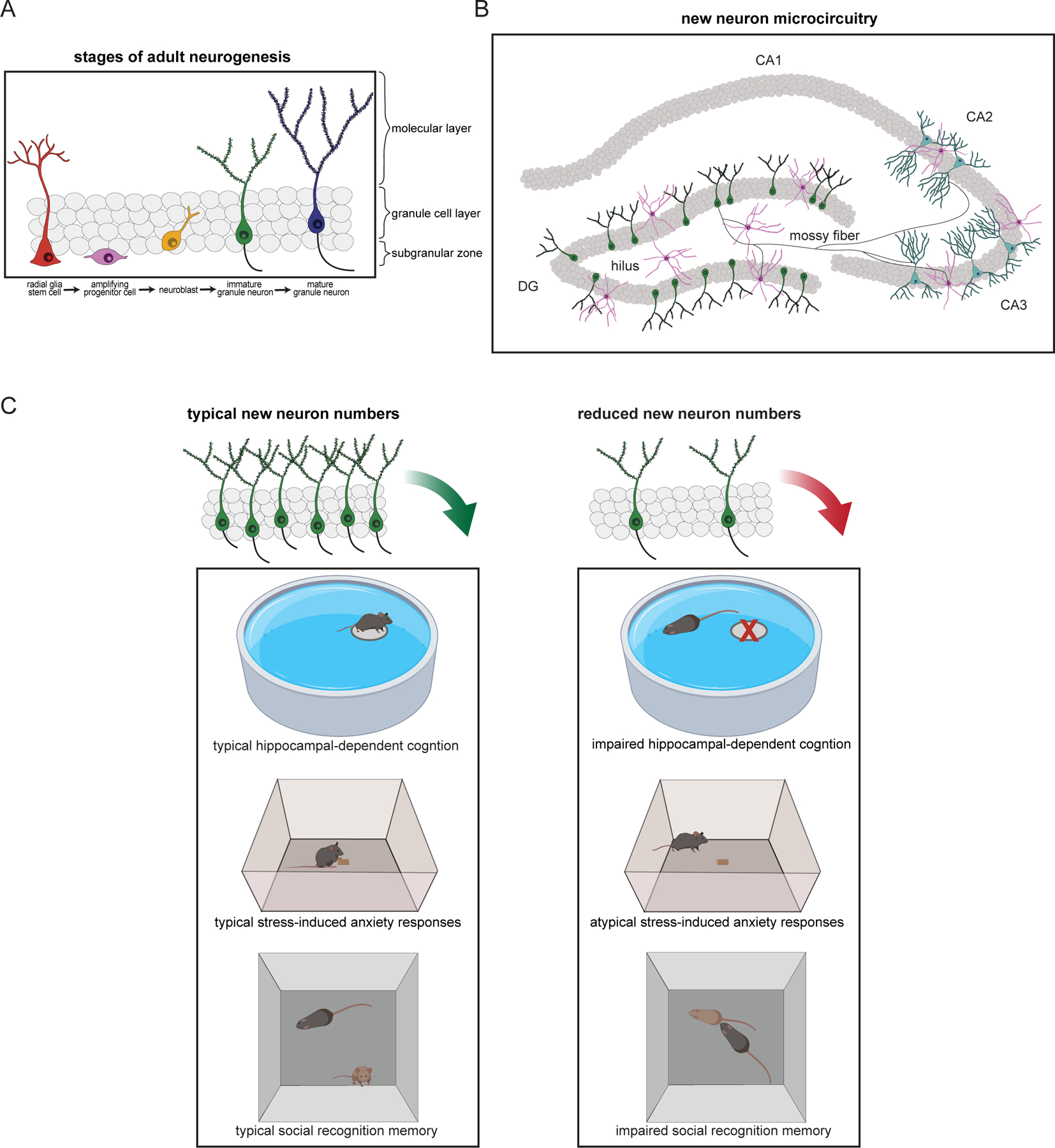
Figure 1. Overview of adult neurogenesis in the hippocampus, the circuitry into which the new cells are incorporated, and their involvement in behavior.

(A) Different stages of adult neurogenesis in the dentate gyrus (DG) of the hippocampus. New granule neurons originate from radial glial stem cells residing in the subgranular zone. These stem cells give rise to amplifying progenitor cells that in turn produce neuroblasts. The neuroblasts migrate into the granule cell layer and develop into immature granule neurons with a single dendritic tree extending toward the molecular layer and an axon (mossy fiber, mf) extending through the hilus toward the CA3 and CA2 subfields. (B) Schematic diagram of new neuron projections, showing the different hippocampal subfields (DG, CA3, CA2, and CA1) which serve as new neuron targets. Mossy fibers from new granule neurons form connections with hilar mossy cells, pyramidal cells in the CA3 and CA2, as well as with inhibitory interneurons in the DG, hilus, and CA3. (C) New granule neurons have been linked to behaviors associated with the hippocampus, such as spatial navigation learning, anxiety/stress regulation, and social cognition. Compared to rodents with typical numbers of new neurons (left), rodents with reduced adult neurogenesis (right) are impaired on some cognitive tests (for example, unable to locate the location of a hidden platform in the Morris water maze), have atypical responses in anxiety tests (for example, different latencies to approach food in the novelty suppressed feeding task), and impaired social cognition (for example, inability to recognize previously encountered mice in a social interaction test).
