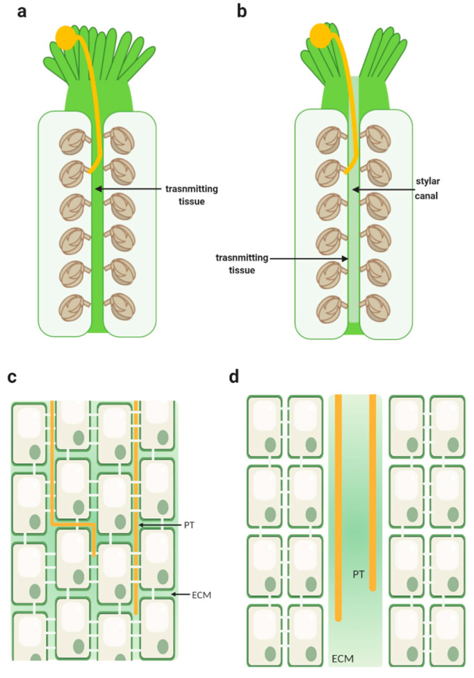
Figure 1.
Schematic representation of a closed and an open style. (a) Longitudinal section of a closed style showing a continuous strand of transmitting tissue inside the pistil. A pollen grain is represented germinating in the stigmatic cells, with a pollen tube (in yellow) growing through the transmitting tract tissue towards the ovules. (b) Longitudinal section of an open style showing a continuous stylar canal lined with a secretory epidermis. A pollen grain is shown germinating in the stigmatic cells, with a pollen tube (in yellow) directing towards the ovules at the surface of the canal cells. (c) Longitudinal section of a part of the transmitting tissue from a closed style exhibiting the presence of substantial intercellular spaces filled with the extracellular matrix. Elongated cells are shown; they are connected through each other by the plasmodesmata in its transverse walls. Pollen tubes (PT) (shown in yellow) are displayed growing between the cells, in the extracellular matrix (ECM). (d) Longitudinal section of an open style showing the epidermal layer of secretory cells lining a canal filled with extracellular matrix (ECM); the pollen tube (PT) is yellow. Created with BioRender.com(accessed on 25 February 2021).

