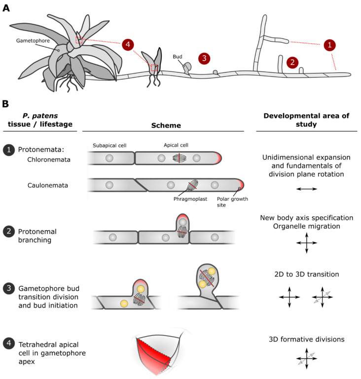
Figure 2.
Developmental progression in Physcomitrium patens and the accompanying cellular phenomena that can be studied. (A) Schematic overview of stages in P. patens (gametophytic) development. Cellular outlines (protonemata/buds) or tissue outlines (gametophores) are depicted. Tissue types predominantly associated with juvenile up to adult phases are arranged right to left. Numbers correspond to particular tissues and/or life stages, with oriented cell divisions leading to tissue type specification or changes in growth axes that are further detailed in B. (B) Four P. patens tissues/life stages where various aspects of cell division plane orientation and the establishment of new organismal axes can be studied: (1) Two types of filamentous protonemata (chloronemata + caulonemata) both grow by polarized, unidimensional cell expansion at their apex. The former produces division planes (red line) perpendicular to the growth axis, while the latter exhibits tilting of the division apparatus (phragmoplast), leading to slanted division planes. (2) A secondary growth axis (indicated by arrows) within protonemal tissue can be established by branching of subapical cells. This involves cell polarization and control over nuclear position and division plane orientation. (3) From the juvenile protonema, a transition to 3D developing gametophores can be initiated. This starts by outgrowth of a bud accompanied by cell divisions with specific division plane orientations that establish new organismal axes. Initiation of this developmental program is regulated by distinct transcriptional and hormonal pathways (indicated by yellow nuclei). (4) The apex of the bud ultimately gives rise to a singular stem cell with three cutting faces (one is indicated). Its continued production of daughter cells and their further developmental trajectories drive gametophore morphogenesis.

