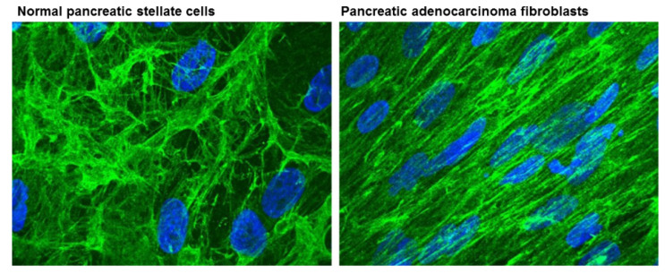
Figure 3.
ECM fiber structures from normal pancreatic stellate cells and pancreatic adenocarcinoma associated fibroblasts stained for anti-fibronectin (green). ECMs organized in parallel patterns were observed in pancreatic adenocarcinoma fibroblasts that are similar to those formed by FAP-positive matrices. In contrast, ECM structures of normal pancreatic stellate cells resemble more FAP-negative matrices. Nuclei were stained with DAPI (blue). Reproduced with permission from [39].

