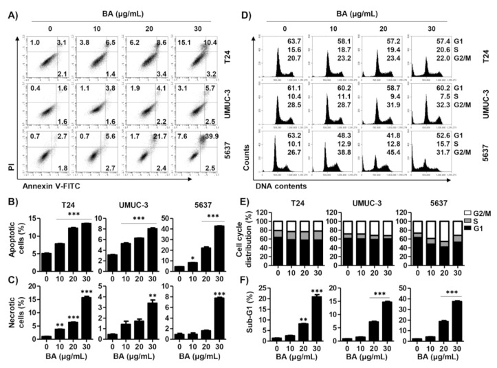
Figure 2.
BA induces apoptosis, necrosis, and cell arrest in human bladder cancer cells. Cells were treated with BA (0, 10, 20, and 30 μg/mL) for 48 h. (A) Cells were collected and stained with annexin V-fluorescein isothiocyanate (FITC)/propidium iodide (PI), and then early apoptotic (annexin V+/PI−), late apoptotic (annexin V+/PI+), and necrotic (annexin V−/PI+) cells were measured by flow cytometry. (B,C) The proportion of apoptotic and necrotic cells were quantified. The percentage of apoptotic and necrotic cells are shown as the mean ± SD (n = 3). ** p < 0.01 and *** p < 0.001 vs. untreated control group. (D) The cell cycle distribution was detected using flow cytometer. (E,F) The percentages of cell cycle distribution and sub-G1 phase cells were quantified. The percentage of cell distribution and sub-G1 phase are shown as the mean ± SD (n = 3). ** p < 0.01 and *** p < 0.001 vs. untreated control group.

