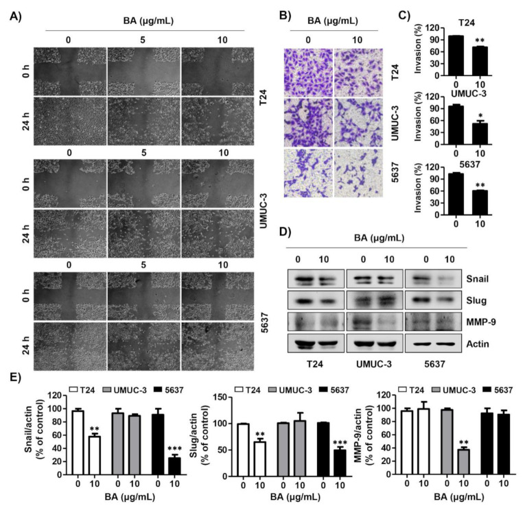
Figure 7.
BA delays migration and invasion of human bladder cancer cells. (A) Cells were seeded, scratched, and then treated with BA (0, 5, and 10 μg/mL) for 24 h. The wound was measured using a phase-contrast microscope (magnification, 50×). (B) Cells were mixed BA with serum free medium and seeded in the upper Trans-well chamber, and then the medium containing 10% fetal bovine serum was added in the lower chamber. After 24 h of incubation, cells were washed, fixed, and stained with 0.1% crystal violet. The invasion cells were observed by a phase-contrast microscope (magnification, 50×). (C) The invading cells were calculated, as compared with the control cells. Data are expressed as the mean ± SD (n = 3). * p < 0.05 and ** p < 0.01 vs. control cells. (D) The expression of migration-associated proteins (Snail, Slug, and matrix metalloproteinase (MMP-9)) were detected using Western blot analysis. Actin was used as a loading control. (E) Bar graphs indicate the relative band density in Western blot analysis (n = 3). ** p < 0.001, and *** p < 0.0001 vs. untreated control group.

