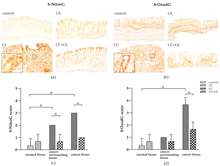
Figure 4.
Immunohistochemical (IHC) staining for (a) 8-NitroG and (b) 8-OxodG in the colonic tissues of the four groups of mice. Brown color indicates specific immunostaining. Cancer surrounding tissue represents the normal cells adjacent to the colon cancer tissue. Original magnification—100×. IHC score for (c) 8-NitroG and (d) 8-OxodG in the colonic tissues of the four groups of mice. Graphs represent the average score (bar: SD; * p < 0.05).

