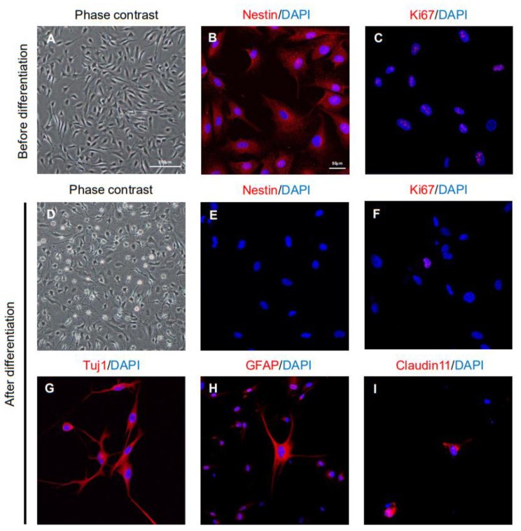
Figure 1.
Characteristics of ahMNCs isolated from patients with hemorrhagic stroke. (A) ahMNCs were shown to be homogeneously bipolar and in the proliferation condition (before differentiation) at in vitro passage 6 (P6). (B,C) ahMNCs at P6 expressed the NSC marker, Nestin (B) and proliferation marker, Ki67 (C). (D) When P6 ahMNCs were subcultured under differentiation conditions (after differentiation), they demonstrated a range of morphological changes associated with differentiated neural cells. (E,F) The expression of Nestin (E) and Ki67 (F) decreased after differentiation. (G–I) Expression of differentiated neural cell markers such as Tuj1 (G, for neuron), GFAP (H, for astrocyte), and Claudin11 (I, for oligodendrocyte) was observed after differentiation. The scale bars = 200 and 50 µm in the phase-contrast and immunofluorescence images, respectively.

