Figure 3.
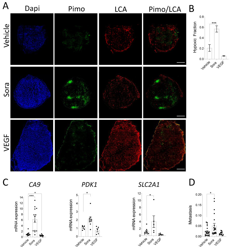
(A–C) HT-1080 cell suspensions were inoculated onto the CAM in the presence or absence of VEGF while Sorafenib (Sora) or DMSO (Vehicle) were added topically to the developing tumors every day. A mix of LCA and pimonidazole was injected into CAM vasculature prior to tumor extraction from the CAM. (A) Representative immunofluorescence images showing regions of hypoxia (pimonidazole; green) and vascularization (LCA; red) in control and treated tumor xenografts are shown. Nuclei were stained with DAPI (blue). Scale bar = 500 µm (B) The hypoxic fraction of control or treated tumor xenografts was quantified (n = 5–7). (C) Relative mRNA expression of CA9, SLC2A1(GLUT1), and PDK1 in treated vs. control tumor xenografts is shown (n = 5–13). (D) Embryonic livers were harvested after 7 days of tumor growth for genomic DNA extraction. The ability of HT1080 cells to disseminate in embryonic livers was quantified as the relative amount of metastasis (Alu) normalized to the amount of host DNA (GAPDH). Metastasis per mm3 of original tumor is shown (n = 12–21). Bars represent the mean ± SEM. The asterisks (*) correspond to p < 0.05 (*) and p < 0.001 (***), one-way ANOVA.

