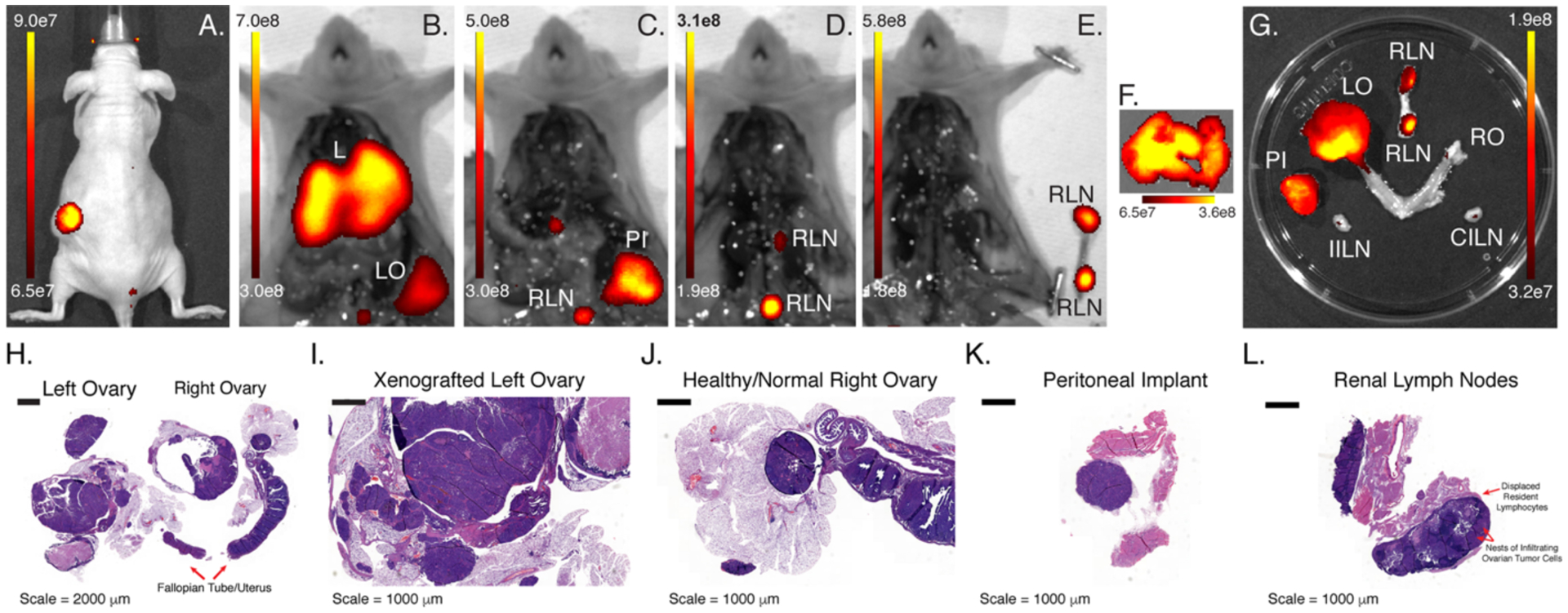
Figure 2. In vivo and ex vivo NIRF imaging using ssB43.13-IR800.

(A) NIRF image of a mouse bearing an orthotopic, CA125-positive OVCAR3 xenograft implanted in the left ovary obtained 120 h after the administration of ssB43.13-IR800 (100 μg; 0.66 nmol); NIRF images obtained after (B) the exposure of the peritoneal cavity, (C) the removal of the liver [L] and left ovary [LO], (D) the removal of the peritoneal implant [PI], and (E) the removal of the renal lymph nodes [RLN]; ex vivo NIRF images obtained of (F) the liver as well as (G) the left ovary, right ovary [RO], renal lymph nodes [RLN], ipsilateral inguinal lymph nodes [IILN] and contralateral inguinal lymph nodes [CILN]; histopathologic analysis of the resected (H) reproductive tract, (I) xenografted left ovary, (J) healthy right ovary, (K) peritoneal implant, and (L) renal lymph nodes
