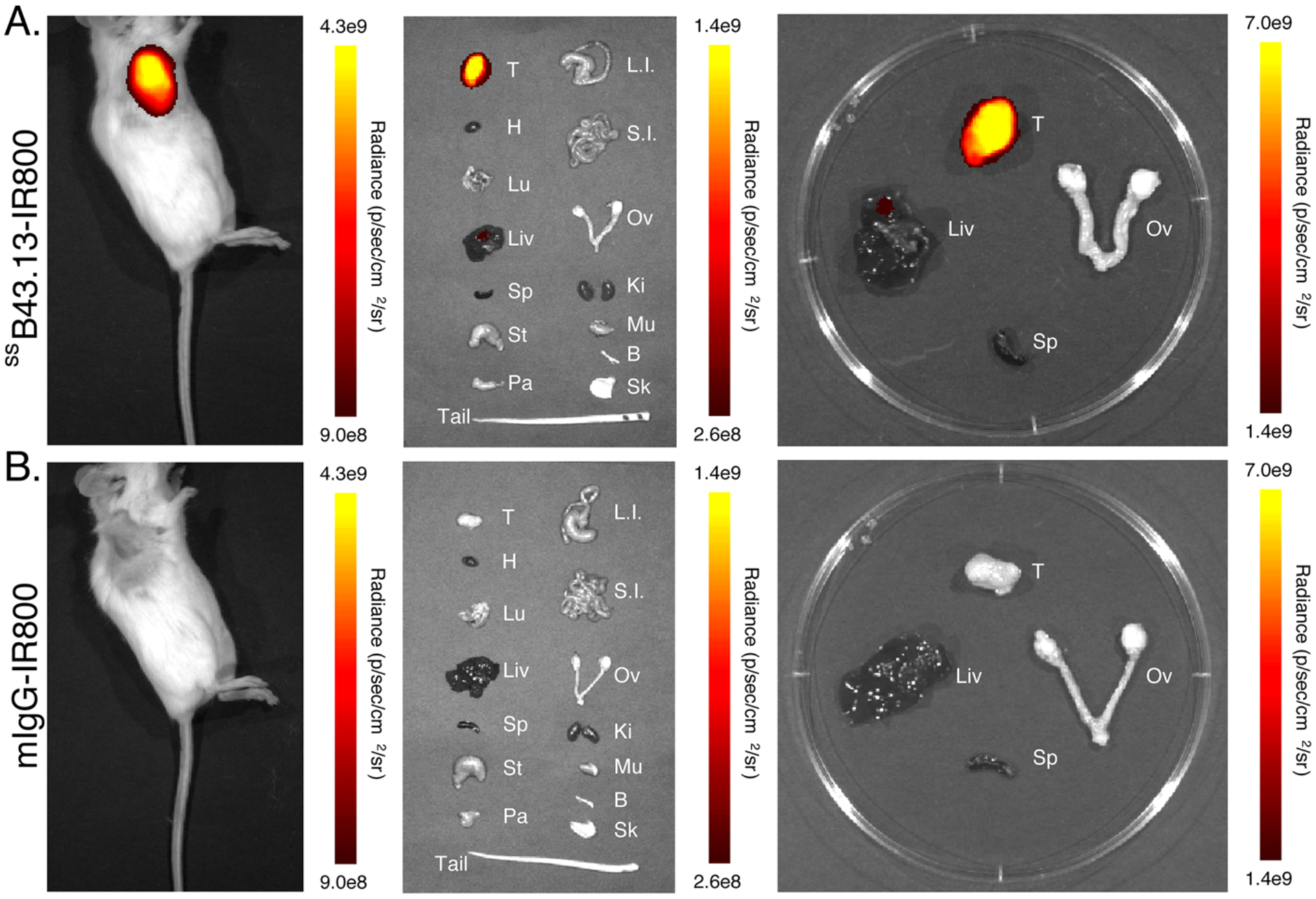
Figure 3. In vivo and ex vivo NIRF imaging with ssB43.13-IR800 and IgG-IR800 in an NSG mouse bearing a subcutaneous OVCAR3 xenograft.

Representative NIRF images were obtained in an NSG mouse bearing a CA125-positive OVCAR3 human ovarian subcutaneous xenograft 72 hours after the intravenous administration of (A) ssB43.13-IR800 (50 μg; 0.33 nmol) and (B) mIgG-IR800 (50 μg; 0.33 nmol). Upon necropsy, selected organs were harvested, and ex vivo NIRF images were obtained: T = tumor; H = heart; Lu = lungs; Liv = liver; Sp = spleen; St = stomach; Pa = pancreas; L.I. = large intestine; S.I. = small intestine; Ov = ovaries; Ki = kidneys; Mu = muscle; Bo = bone; Sk = skin.
