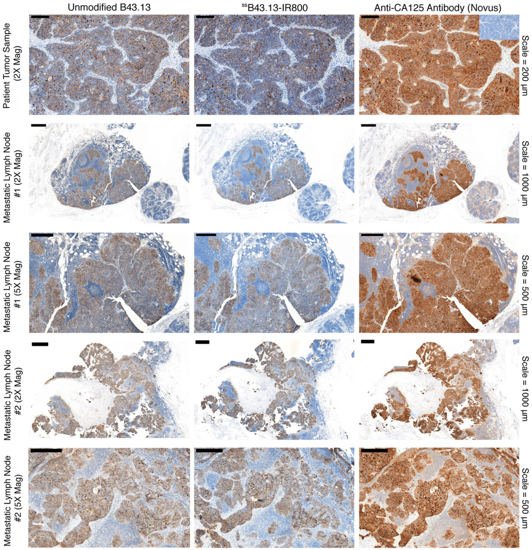
Figure 5. Histopathologic analysis of tumor and lymph node samples from a patient with metastatic HGSOC.

Immunohistochemical staining of CA125 in tumor and lymph node samples obtained from a patient who underwent surgical debulking to remove peritoneal tumor masses and metastatic lymph nodes at Memorial Sloan Kettering Cancer Center. The inset image on the top right corner shows staining of the HGSOC tumor section with mouse isotype IgG1 antibody. A high concordance was observed in the staining patterns (brown) when equivalent dilutions of unmodified B43.13, ssB43.13-IR800, and a commercially available anti-CA125 antibody (Novus Biologicals, NBP1–96619) were employed for immunohistochemistry. IHC images are representative of samples from 3 independent HGSOC patients.
