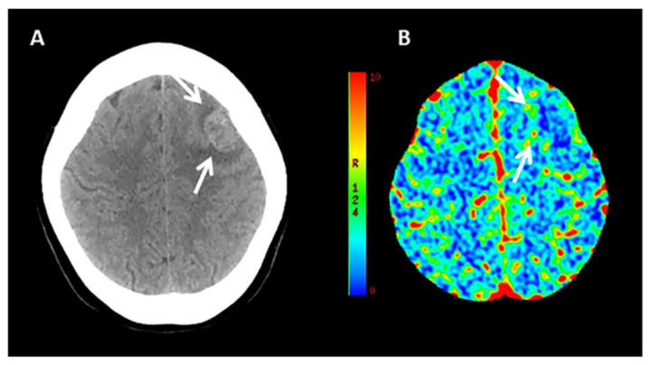
Figure 1.

Example of CT Perfusion in a patient with ICH due to malignancy. Non-contrast CT (NCCT) scan (A) and cerebral blood volume (CBV) map (B) in a patient with acute ICH due to the bleeding of an underlying high grade glioma located left frontal lobe and characterized by elevated CBV values in the perihematomal region (arrows).
