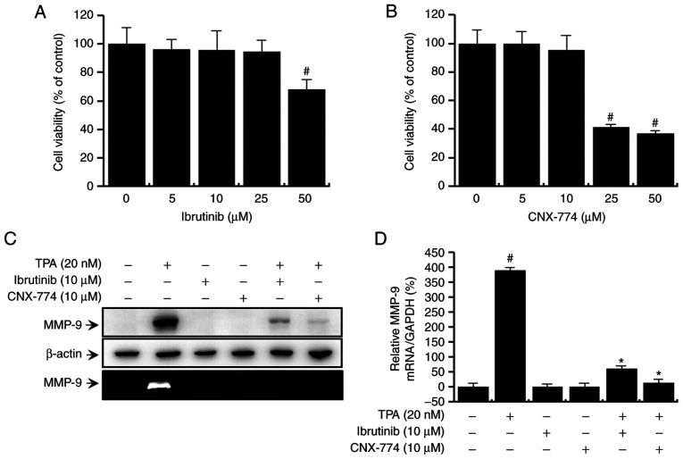
Figure 1.
Effect of BTK inhibitors on the MCF-7 cells by TPA-induced MMP-9 expression. To examine the cytotoxicity of BTK inhibitors, cells were cultured in 96-well plates to 70% confluency and various concentrations of (A) ibrutinib or (B) CNX-774 were added to the cell culture and incubated for 24 h. The EZ-Cytox Enhanced Cell Viability Assay Kit was used to determine cell viability. The optical density of the control was considered 100%. Data represent the mean ± SEM of three independent experiments. #P<0.05 compared with 0 µM of BTK inhibitors at 24 h. MCF-7 cells were treated with BTK inhibitors in the presence or absence of TPA for 24 h. Cell lysates were analyzed by western blotting using an anti-MMP-9 antibody. The blot was re-probed with an anti-β-actin antibody to confirm equal loading. Conditioned medium was prepared and used for gelatin zymography (C). MMP-9 mRNA levels were analyzed by quantitative PCR and GAPDH was used as an internal control Data are the mean ± SEM of three independent experiments. #P<0.01 vs. untreated control; *P<0.01 vs. TPA. (D) The blot was re-probed with an anti-β-actin antibody to confirm equal loading.

