Fig. 1.
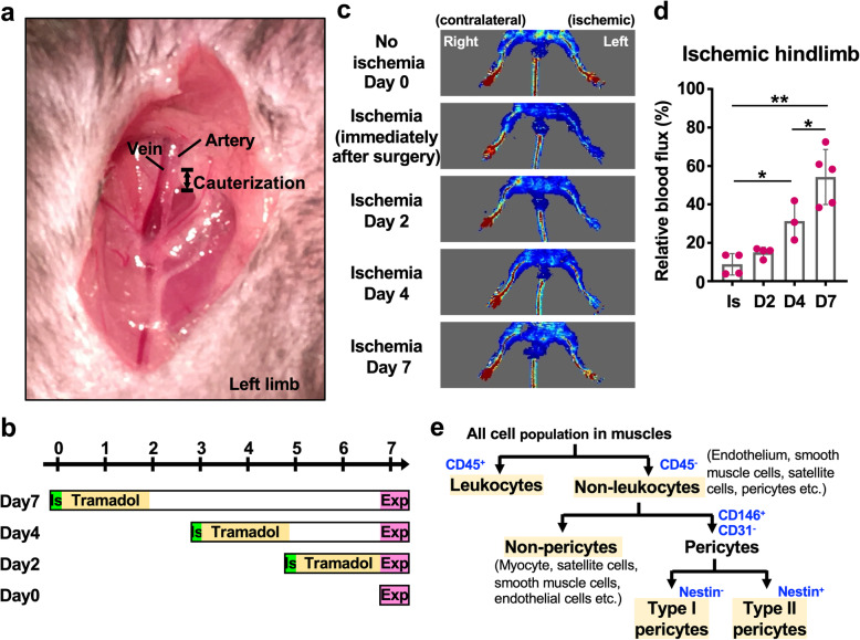
Analysis of pericyte biology in a mouse model of hindlimb ischemia. a Surgery to induce hindlimb ischemia was performed on the left leg of the mouse. After dissociating the artery from the vein, a segment of the artery was electrocauterized. b Schematic graph of the timeline for sample collection after ischemia surgery. IS, ischemia; Exp, experiment (i.e., flow cytometry and FACS). c The Doppler images show the blood flow in the ischemic and contralateral limbs at 0, 2, 4, and 7 days after surgery. The red signal represents the abundant blood flow. d The quantitation of blood flux of ischemic hindlimbs (left) relative to contralateral hindlimbs (right). The measurement was performed by MoorLDI software (version 5.2, Moore Instruments, Axminster, UK). Student’s t test was used to perform statistical analyses (*p < 0.05; **p < 0.01). IS, immediately after ischemia surgery. The mouse numbers used for Doppler scanning on ischemia day 0, 2, 4, and 7 were 4, 4, 3, and 5, respectively. e The strategy used to identify endothelial cell and pericyte populations in flow cytometry and FACS experiments

