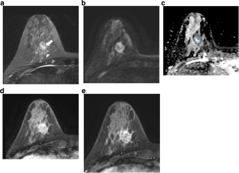
Fig. 1.
Images of a 37-year-old woman with invasive ductal carcinoma and a lymphovascular invasion. a Axial T2 weighted image shows a 2.3 cm mass in her right breast. Intratumoral T2 high signal (arrow) and peritumoral edema (arrowhead) are noted. b High signal intensity rim is shown in axial diffusion weighted image (b value=1000 s/mm2). c Apparent diffusion coefficient map shows restriction along the periphery of the mass. Multiple region of interests (ROI)s of 6.87 mm2 were manually placed within the mass avoiding cystic or necrotic area. Minimum ADC value was 1025 × 10−6 mm2/s, and maximum ADC value was 1345 × 10−6 mm2/s. The calculated ADC difference was 320 × 10−6 mm2/s. d, e Axial contrast-enhanced T1-weighted image after 2 min (d) and 6 min (e) of contrast injection demonstrating a round, circumscribed mass in right breast. This mass shows rim internal enhancement and washout kinetic pattern of enhancement. This patient underwent modified radical mastectomy of her right breast. The histopathological features of this mass were poorly differentiated, no lymph node metastasis, ER-. PR-, HER2- and Ki-67 high (70%). Tumor size at final pathologic report was 3.6 cm

