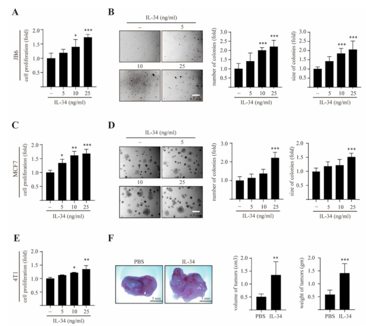
Figure 1.

Effects of IL-34 on anchorage-independent growth and mammary gland tumorigenesis in vitro and in vivo. (A) JB6 Cl41 cells were treated with different concentration of IL-34 for 48 h, following which the cell proliferation was estimated using BrdU incorporation assay. (B) JB6 Cl41 cells were exposed to different concentrations of IL-34 as indicated, subjected to soft agar assay, and incubated at 37 °C in a 5% CO2 atmosphere for 14 days. Representative colonies from three separate experiments were photographed (left), followed by calculation of the average colony numbers and sizes (right). (C) MCF7 cells were treated with various doses of IL-34 for 48 h and cell proliferation was approximated using BrdU incorporation assay. (D) MCF7 cells were treated with the indicated concentration of IL-34 in a soft agar matrix and incubated at 37 °C in a 5% CO2 atmosphere. After 14 days, colonies from three separate experiments were photographed (left), followed by calculation of the average colony number and sizes (diameter > 100 μm, right). (E) 4T1 cells were seeded and treated with different concentrations of IL-34 for 48 h. Cell proliferation was then estimated using BrdU incorporation assay. (F) 4T1 cells were injected into the mammary gland of BALB/c mice in the presence or absence of 100 ng/mL IL-34 and allowed to grow until tumors were formed. Shown are representative pictures of tumor (left), measured volumes and weights of tumors (right). Error bars indicate mean ± S.D. of triplicate measurements from three independent experiments. Statistical analyses were conducted using one-way ANOVA (*p < 0.05, ** p < 0.01, *** p < 0.001, compared to the control groups).
