
Figure 2. Cryo-EM reconstruction of the human PIKfyve complex at medium- low resolution.
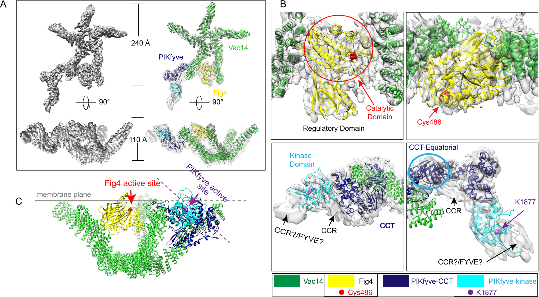
(A) The composite reconstruction alone and with model. The left panels show the maps only; the right panels also show docked protein models colored according to panel 1A. (B) Enlarged views showing the fit of the Fig4 Sac homology module (top panels) and the PIKfyve CCT and kinase modules (bottom panels) to the map. Two different views for Fig4 and PIKfyve are related by 90° rotations. (C) Model for PIKfyve complex interacting with membrane. The Fig4 active site is oriented to face the membrane, whereas the PIKfyve active site is twisted away from the membrane by ~45° and so cannot access membrane incorporated phosphoinositides.