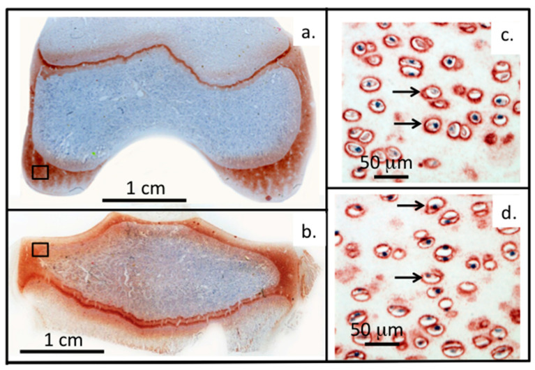
Figure 2.
Perlecan and FGF-2 are prominent components of the PCM of chondrocytes. (a–d) Immunolocalisation of perlecan in an adult ovine stifle joint. Left panel: Macroscopic views showing femoral condyle (a) and tibial plateau (b). Right panel: High power views demonstrating perlecan’s pericellular distribution in femoral condyle (c) and tibial plateau (d). Figure (a–d) based on data originally published in [50].

