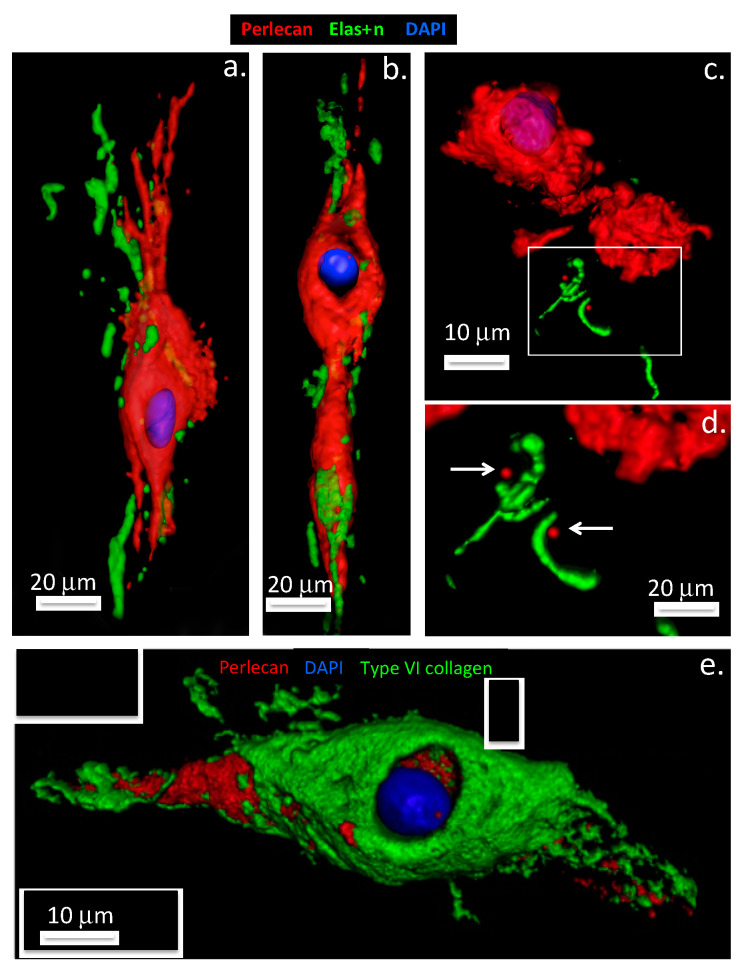
Figure 3.
Perlecan promotes elastin formation by IVD cells. Three-dimensional surface rendered models of perlecan, elastin and type VI collagen immunolocalization in intervertebral disc tissues, based on confocal data originally published in [54,38]. Upper panel: Annulus fibrosus cells shown in (a,b), nucleus pulposus cells in (c,d). Secretion of co-acervated tropoelastin (green) is visible at the poles of the elongate, spindle shaped annulus fibrosus cells, perlecan label (red) extends outwards from the pericellular matrix compartment at the cell poles (a,b). In contrast, elastin is only sparsely distributed around NP cell clusters (c,d). Detail of the elastin labeling (boxed area in (c) is shown in (d) Arrows denote perlecan foci associated with elastin label in (d). Lower panel: The lower image (e) shows the overlapping distributions of collagen type VI (green) and pericellular perlecan label (red) within an annulus fibrosus chondron. Cell nuclei depicted in blue.

