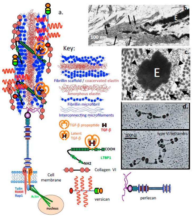
Figure 4.
Schematic depiction of the elastic microbril, containing perlecan and fibrillin as functional omponents. (a) Schematic depiction of an elastic microfibril and its components. Electron micrographs of tannic acid-glutaraldehyde-fixed samples demonstrating the central amorphous elastin (E) within a microfibril in (b) longitudinal (magnification ×18,000) and (c) cross-section (magnification ×85,000); (d) Rotary shadowing images showing the characteristic interrupted globular domains in type VI collagen. Segment b, c reproduced from [55]. Segment d [56] under Open Access. Arrows in (b). depict microfibrillar material, E amorphous elastin. Arrowheads in (c). depict microfibrils in cross section, whilst arrows indicate collagen fibres in cross section.

