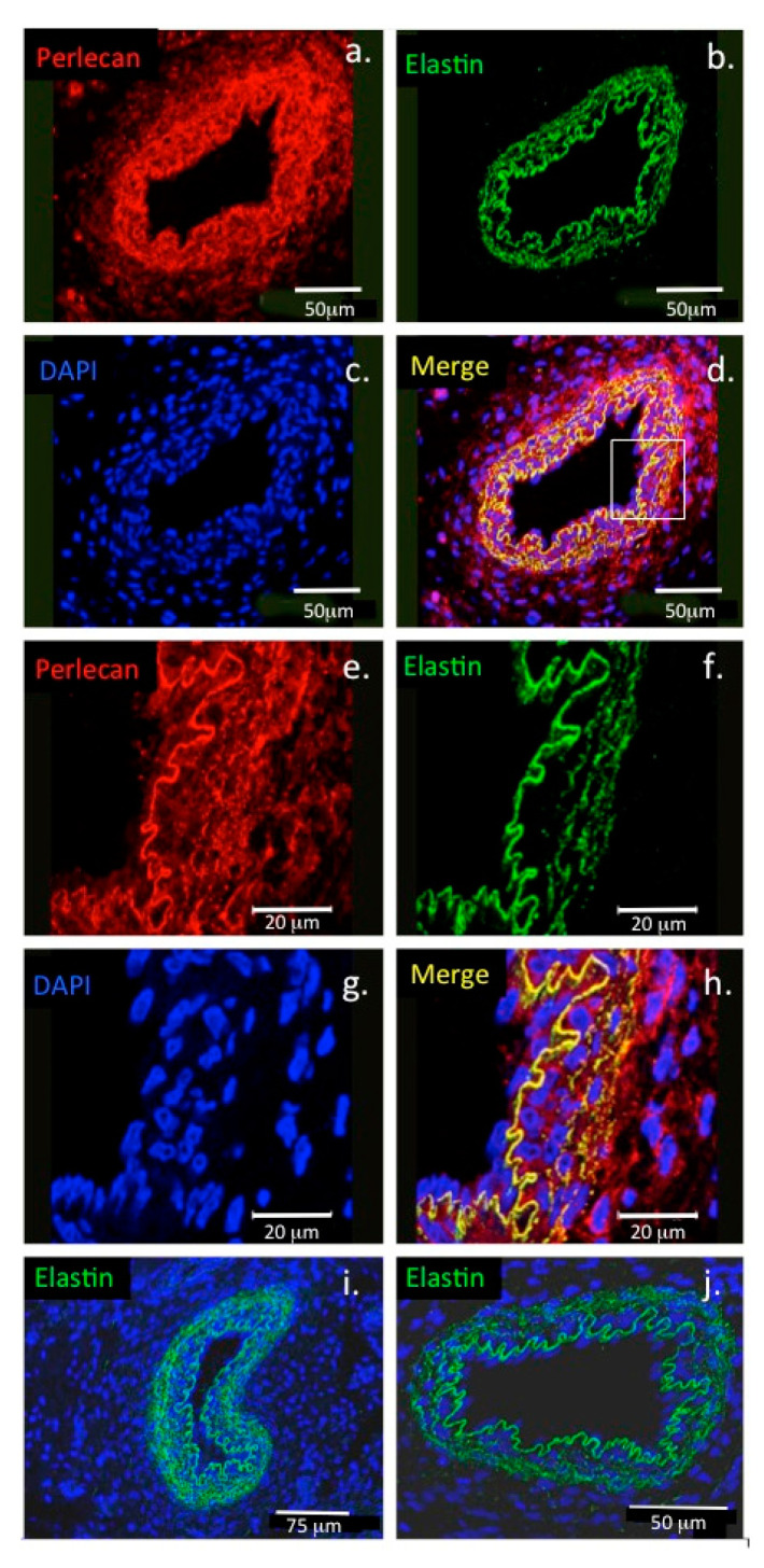
Figure 6.
Colocalisation of perlecan and elastin in blood vessels. Confocal immunofluorescence localisations of perlecan and elastin in the basement membrane of a paraspinal blood vessel from a 14-week-old human foetal spinal specimen (a–d). Perlecan (red) and elastin (green) are colocalised (yellow) at the lumenal surface of the endothelial cell basement membrane of the blood vessel. The boxed area in (d) is shown at higher magnification in (e–h). (i,j) Elastin immunolocalisations are also shown in an additional paraspinal capillary (i) and in a venule (j). Cell nuclei depicted with DAPI staining (blue). Image reproduced from [54].

