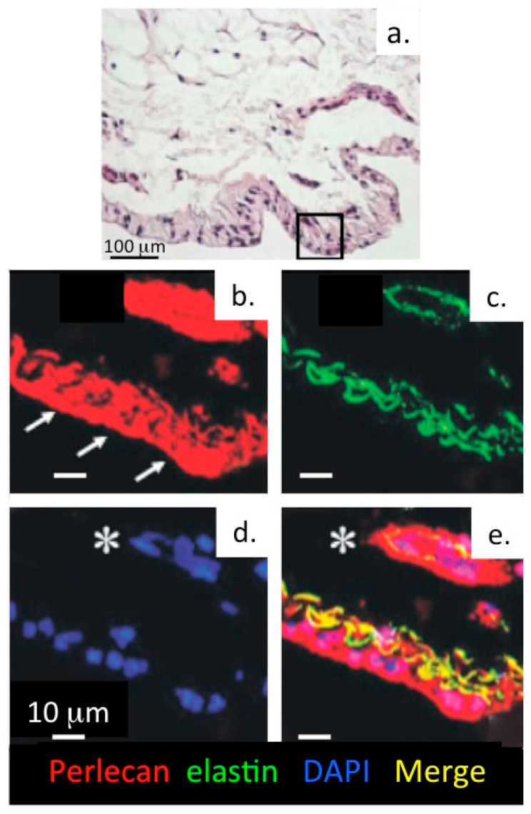
Figure 8.
Perlecan and elastin are components of a basal lamina in the knee joint endothelium. (a) Haematoxylin & eosin stained tissue section of an 18 month old ovine knee synovium specimen. Confocal localisation of perlecan (b, red), elastin (c, green), DAPI-stained cell nuclei (d) and merged image showing colocalisation of perlecan and elastin (e, yellow) within an elastic lamina and surrounding a synovial blood vessel. The arrows indicate the surface of the synovium.The asterisk indicates a blood vessel in the synovium. Figure reproduced from [54] with permission.

