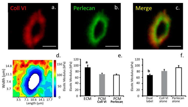
Figure 10.
Correlative co-localization of perlecan and type VI collagen with the biomechanical properties of cartilage. (a–c) Dual immunofluorescence labeling of type VI collagen (a, red), and perlecan (b, green) within the pericellular matrix compartment of articular cartilage. The adjacent overlay image (c) shows their overlapping distributions. Scale bar = 5 μm. (d) Contour map of the calculated elastic moduli for the pericellular matrix region shown in (a–c), as determined by AFM scanning. (e) Elastic moduli of cartilage extracellular matrix (ECM) and pericellular matrix (PCM) as defined by the presence of type VI collagen or perlecan. There is no difference between biochemical definitions of the PCM (p = 0.70). ECM elastic moduli were significantly greater than PCM moduli (a: p < 0.005 as compared to either PCM definition). (f) Regions that were dual-labeled for type VI collagen and perlecan exhibited lower elastic moduli than regions positive for type VI collagen or perlecan alone (b: p < 0.05 as compared to type VI collagen and perlecan alone regions). Data presented as Mean + SEM. Figure reproduced from [43] with permission.

