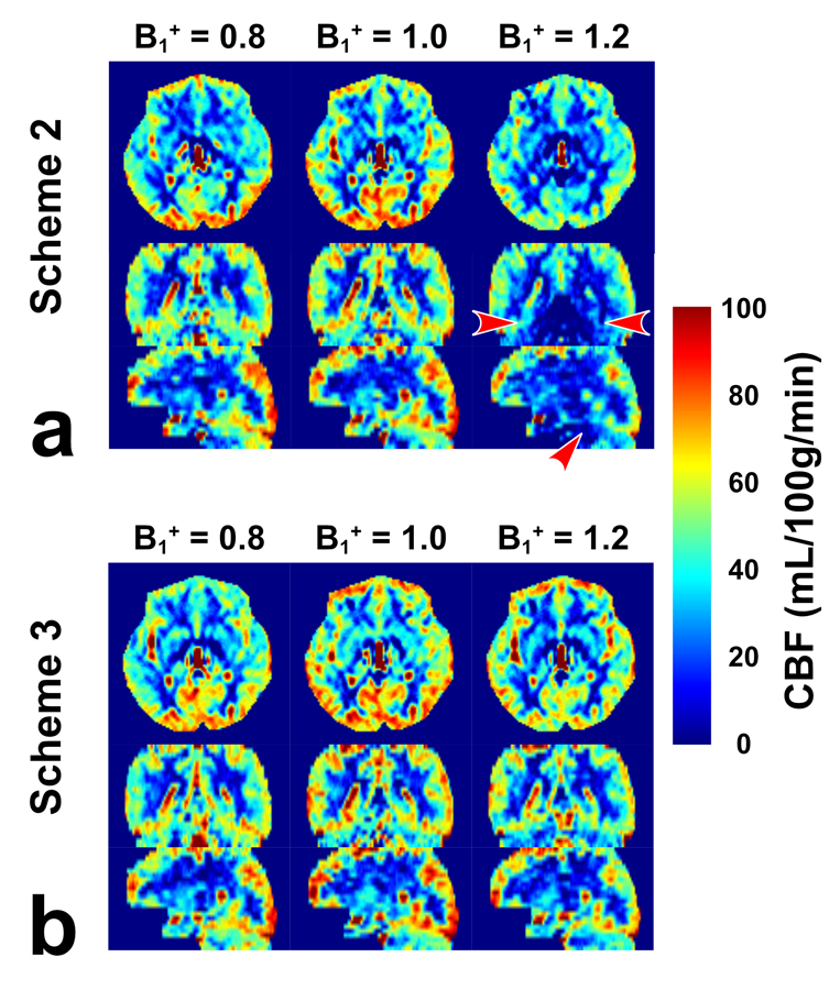
Figure 6:

CBF maps acquired from the FT-VSI prepared brain ASL along axial, sagittal and coronal views at the B1+ scale of 0.8, 1.0, and 1.2 with (a) scheme 2 using velocity-compensated control and (b) scheme 3 using dynamic phase-cycling. Scheme 3 markedly reduced the pseudo perfusion deficit at the base of the brain produced by scheme 2 with the B1+ scale of 1.2 (a, red arrowhead).
