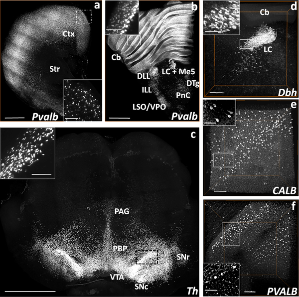
Fig. 4. Volume rendered visualizations of Pvalb, Dbh, and Th mRNA expressing neurons in the rat brain, and CALB+ and PVALB+ neurons in the postmortem human cerebellum following HCR FISH and iDISCO+.
(a) COLM-acquired volume of Pvalb expressing neurons in a 2 mm thick rat cortex (Ctx) and striatum (Str) hemi-slice between bregma = 2.00 mm and 0.00 mm. (b) COLM-acquired image of Pvalb expression pattern in a 2 mm rat brainstem hemi-slice between bregma = −8.44 mm and −10.44 mm. (c) COLM-acquired image of Th expression in a 2.5 mm thick rat mid-brain volume between bregma = −4.40 mm and −6.90 mm. (d) Confocal microscope-acquired image stack of Dbh expression in the locus coeruleus (LC) of rat brainstem hemi-slice between bregma = −9.72 mm and −10.32 mm. (e) CALB and (f) PVALB expression in the postmortem human cerebellum of a control subject (#2292) acquired on confocal microscope (1200 μm deep stacks). Cb- cerebellum, DTg- dorsal tegmental nucleus, ILL/DLL- intermediate/dorsal lateral lemniscus, LC+Me5- locus coeruleus + mesencephalic trigeminal nucleus, PnC- caudal pontine reticular nucleus, LSO/VPO- lateral/ventral superior olive, PAG- periaqueductal gray, PBP- parabrachial pigmented nucleus of the VTA, SNc/r- substantia nigra compacta/reticulata, VTA-ventral tegmental area. Scale bars (μm) - (a-b) 1000 (Inset-400); (c) 500 (Inset-100); (d) 1000 (Inset-200); (e-f) 500 (Inset-250).

