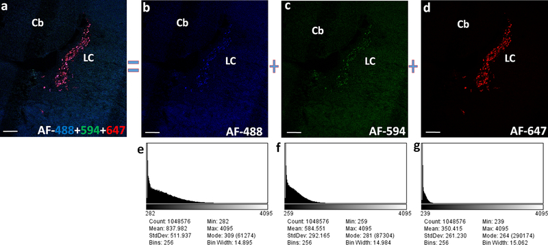
Fig. 6. Fluorophore based differences in the fluorescence output imaged using confocal microscope.
(a) Dbh expression in the rat locus coeruleus (LC) following multiplexed HCR FISH using a set of common mRNA binding probes and 3 different hairpins AF-488, AF-594 and AF-647. Relatively weak fluorescence and high background recorded with the use of AF-488 (b) and AF-594 (c) tagged DNA hairpins in comparison to the strong fluorescence with low background following the use of hairpins conjugated with AF-647 (d). Screenshots of the histogram (e-g) for their respective z-projection images (b-d), generated in the ImageJ software depict the poor S/N ratio resulting from the use of AF-448 and AF-594 compared to a better S/N ratio from the use of AF-647. Images are pseudocolored for the overlaying purpose. Scale bars- 200 μm.

