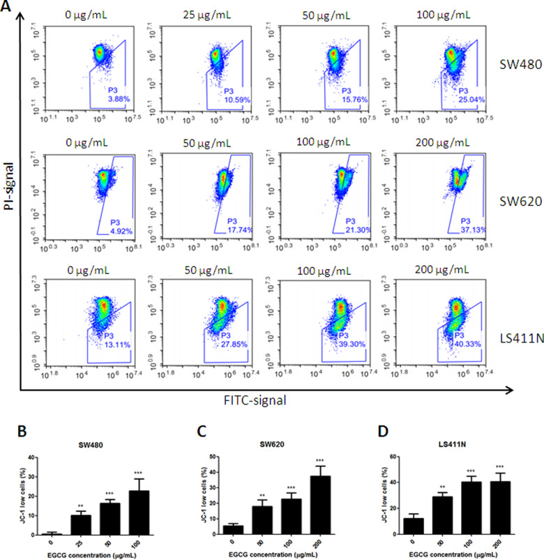
Figure 3.
Collapse of the mitochondrial-membrane potential induced by EGCG. (A) Flow-cytometry images. (B)–(D) Quantitative analysis of the percentage of JC-1 low cells in SW480 (B), SW620 (C), and LS411N (D) cell lines, after treatment with EGCG. The percentages represent cells with a depolarized mitochondrial membrane. **P < 0.01 and ***P < 0.001, as compared with untreated control.

