
Figure 1. Ultrawide-field (UWF) fluorescein angiogram (FA) and fundus photographs of a representative treatment-naïve eye with proliferative diabetic retinopathy (PDR) before and after panretinal photocoagulation (PRP).
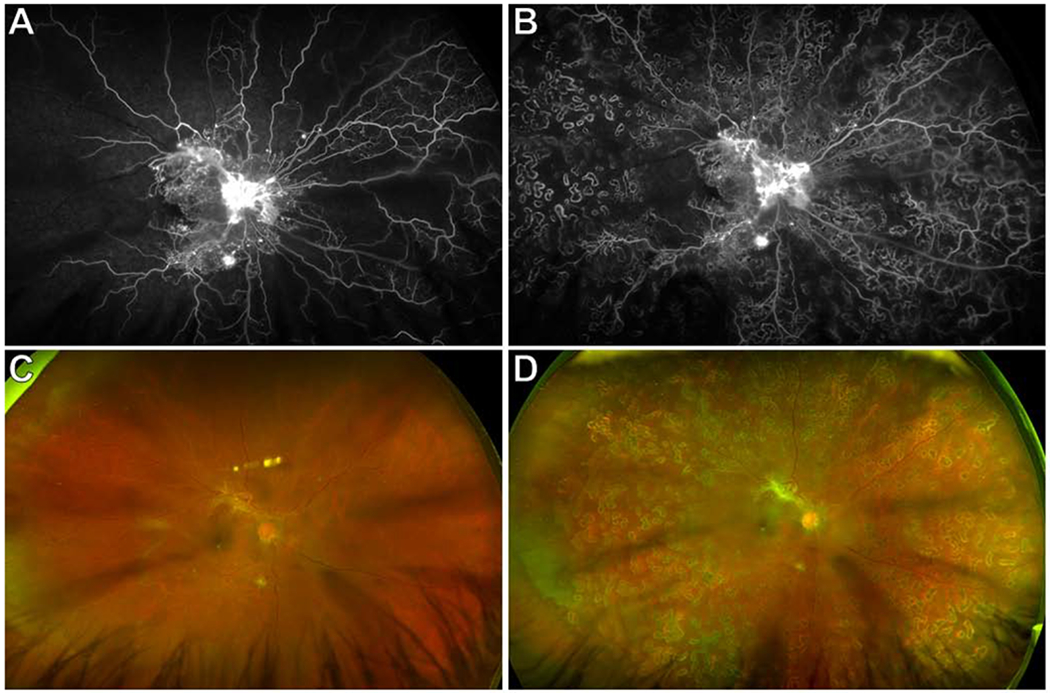
(A) Baseline UWF FA showed neovascularization (NV) of the disc, multifocal NV elsewhere (NVE), and extensive retinal non-perfusion (RNP) involving the macula. Areas of RNP were only evident as “featureless fundus” on the corresponding UWF fundus photograph (C). There was a cortical cataract.
(B) UWF FA 3 months after PRP showed 360 degree dense PRP scars, persistent NVD and NVE, and stable RNP. The corresponding UWF fundus photograph is shown in (D).