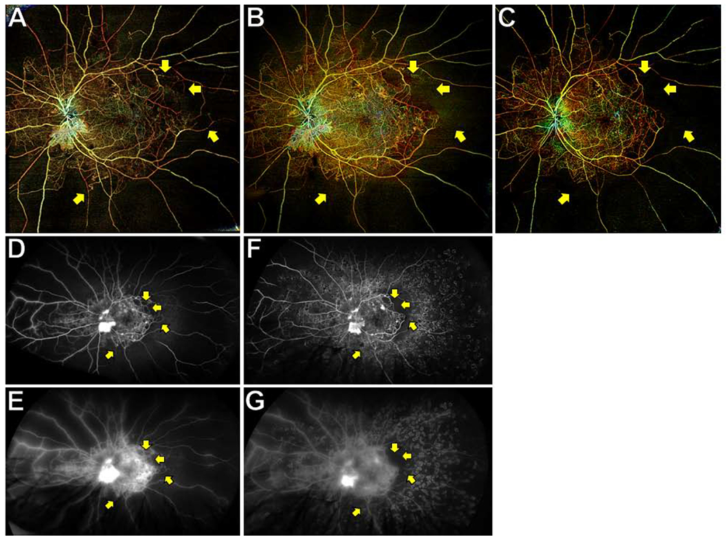
Figure 3. Wide-field (WF) swept-source (SS) OCT angiography (OCTA) posterior pole montages and ultrawide-field (UWF) fluorescein angiograms (FA) of a representative eye with proliferative diabetic retinopathy (PDR) after panretinal photocoagulation (PRP).

WF SS-OCTA posterior pole montages of total retinal vasculature at the baseline (A) and 3 month (B) visits showed retinal non-perfusion (RNP) that corresponded with UWF FAs taken on the same visits (D and E are early and late frames of baseline FA; F and G are early and late frames of 3 month FA). RNP was easier visualized on the 3 month OCTA image (B) than the 3 month FA because of staining of PRP scars (F). RNP was stable at the 3 month and 6 month (C) visits, though some small areas of large-caliber vessel dropout were noted (yellow arrows).
